NucMM dataset: 3d neuronal nuclei instance segmentation (Paper)
Problem description
The goal is to segment and identify automatically each cell nuclei in EM images. To solve such task pairs of EM images and their corresponding instance segmentation labels are provided. Below a pair example is depicted:
MitoEM-H tissue image sample. |
Its corresponding instance mask. |
In this dataset 27 3D images of size (64, 64, 64) voxels, for (z,x,y) axes, are used for train while the test is
done over the whole Zebrafish volume. Here is a training sample and its ground truth:
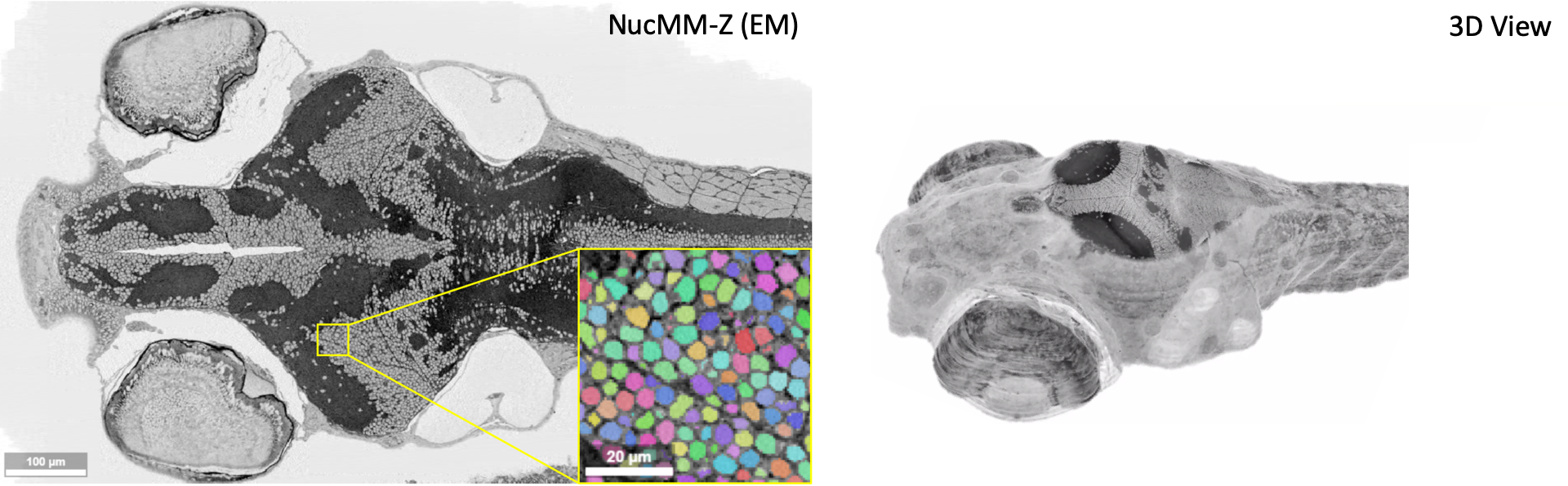
Overview of the NucMM-Z dataset volume. Electron microscopy (EM) volume covering nearly a whole zebrafish brain. Modified image from [8].
Data preparation
You need to download NucMM dataset first from this link. Once you have donwloaded this data you need to create a directory tree as described in Data structure. To adapt the .h5 file format provided by MitoEM authors into .tif files you can use the script h5_to_tif.py.
Configuration file
To create the YAML file you can use the template 3d_instance_segmentation.yaml which is prepared for this tutorial.
How to run
To run it via command line or Docker you can follow the same steps as decribed in the How to run section of the instance segmentation workflow documentation.
Results
The results follow same structure as explained in the Results section of the instance segmentation workflow documentation. The results should be something like the following:
The resulting instance segmentation should be something like this:
Instance segmentation results on the whole dataset.
Zoom of a small region of the instance prediction.